NICK TRANSLATION KIT: PREPARING THE REAGENTS

NICK TRANSLATION KIT image

NICK TRANSLATION KIT: PREPARING THE REAGENTS

0.2 mM SpectrumGreen, SpectrumOrangeTM or 

Add 10 µL of 1 mM dUTP to 40 µ nuclease-free water.


0.1 mM dTTP

Add 10 µL of 0.3 mM dTTP to 20 µL nuclease-free water.


0.1 mM dNTP mix

Mix together 10 µL each of 0.3 mM dATP, 0.3 mM dCTP, and 0.3 mM dGTP.


Extracted P1, BAC, or YAC DNA

Prepare a 0.2µ g/µ to 1µ g/µ solution of extracted DNA in Tris-EDTA (10 mM Tris, 1 mM EDTA, pH 8.5) buffer. 

NICK TRANSLATION KIT: ASSAY PROCEDURE

This procedure labels approximately 1 µg of extracted P1, BAC, or YAC DNA. This is enough for ten FISH experiments (one target area equal to 22 mm x 22 mm). 

  1. Place a microcentrifuge tube on ice and allow the tube to cool.
  2. Add these components to the tube in the order listed. Briefly centrifuge and vortex the tube before adding the enzyme (last component):
    nick-translation-assay-procedure-chart.gif
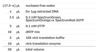
  3. Briefly centrifuge and vortex the tube.
  4. Incubate 8 - 16 hours at 15 °C.
  5. Stop the reaction by heating in a 70 °C water bath for 10 minutes.
  6. Chill on ice.

Determining the Probe Size

Determining the probe size is an essential part of the labeling procedure. For detailed instruction on preparing and running an agarose gel see (1) Maniatis T, Fritsch EF, and Sambrook J. Gel electrophoresis of DNA. In: Molecular cloning: a laboratory manual. 2nd ed. Cold Spring, NY: ColdSpring Harbor Laboratory; 1989 or (2) Ausubel FM, ed. Preparation and Analysis of DNA. In: Current Protocols in Molecular Biology. New York: Greene Publishing Associates and John Wiley & Sons, 1989.


NOTE: Because of the isomeric structures of the fluorophores, the unincorporated dUTP appears on the agarose gel as two domains of differing brightness. The chemical structure and net charge of the different fluorophores affect the migration pattern on the gel. For example, unincorporated SpectrumGreenTM dUTP migrates to the top of the gel, while SpectrumOrangeTM and SpectrumRedTMdUTP migrate further down the gel and may overlap the probe DNA smear.In this case,you need to remove the unincorporated dUTP by ethanol precipitation and/or gel filtration of the labeled probe DNA before loading a sample of the probe DNA on the agarose gel for sizing.


As you increase the amount of enzyme and the incubation time, the size distribution shifts to progressively smaller probe fragments. To produce smaller probe fragments use the following conditions that are listed in order of decreasing fragment size:


5 µL enzyme mix/8 hour incubation, 5 µL enzyme mix/16 hour incubation, 10 µL enzyme mix/8 hour incubation and 10 µL enzyme mix/16 hour incubation. Adjust the amount of nuclease-free water added to keep the total reaction volumeat 50 µL.

PERFORMING FISH WITH THE LABELED PROBE

The following is a recommended protocol for hybridization of unique sequence probes to lymphocyte cells. You may need to modify the procedure depending on the type and size of the probe.


Precipitating the Probe

  1. Pipet 5 µL (~100 ng of probe) of the nick translation reaction mixture into a microcentrifuge tube.
  2. Add 1 µg COT-1 DNA, 2 µg human placental DNA and 4 µL purified water to the tube.
  3. Add 1.2 µL (0.1 volume) 3 M sodium acetate, then add 30 µL (2.5 volumes) of 100% EtOH to precipitate the DNA. Vortex briefly and place on dry ice for 15 minutes.
  4. Centrifuge at 12,000 rpm for 30 minutes at 4 °C to pellet the DNA.
  5. Remove the supernatant and dry the pellet for 10 - 15 minutes under a vacuum at ambient temperature.
  6. Resuspend the pellet in 3 µL purified water and 7 µL Hybridization Buffer.
  7. Denature the probe by heating the probe mix for 5 minutes in a 73 °C water bath.

Hybridizing the Probe to the Target Metaphase

  1. Mark hybridization areas on the slide using a tipped scribe.
  2. Ensure the temperature of the denaturation solution (70% formamide in 2X SSC, pH 7.0 - 8.0) is at 75 °C. Immerse the slide containing normal metaphase spreads into the solution for 5 minutes.
  3. Dehydrate the slide for 1 minute in 70% EtOH, followed by 1 minute in 85% EtOH, and 1 minute in 100% EtOH.
  4. Dry the slide by touching the bottom edge to a blotter and wiping the underside with a paper towel.
  5. Place the slide on a 45 - 50 °C slide warmer to allow remaining EtOH to evaporate.
  6. Apply 10 µL of denatured probe mix to the slide.
  7. Immediately apply the coverslip and seal with rubber cement.
  8. Place slide in a sealed, humidified box and place in a 37 °C incubator for 16 hours for hybridization.

Washing the Slide

  1. Place the container that has the 0.4X SSC/0.3% NP-40 wash solution in a73 ± 1 °C water bath for at least 30 minutes prior to use. Discard after using 1 day. Prepare a second container of 2X SSC/0.1% NP-40 atambient temperature.
  2. Remove the rubber cement seal and the coverslip and immediately place the slide into the 0.4X SSC/0.3% NP-40 wash solution at 73 ± 1 °C. Agitate the slide for 1 - 3 seconds.
  3. Repeat step 2 for each slide - do not wash more than four slides at once - and then let the slides stand for 2 minutes.
  4. Place the slide in the 2X SSC/0.1% NP-40 wash solution at ambient temperature. Agitate the slide 1 - 3 seconds and then let stand for 5 seconds to 1 minute.
  5. Air dry slide in darkness.

Visualizing the Hybridization

Apply 10 µL of DAPI II counterstain and a coverslip to each hybridization location.