Urovysion Kit Contents
The UroVysion Bladder Cancer Kit probes are directly labeled with one of the Vysis fluorophores; SpectrumRed, SpectrumGreen, SpectrumAqua or SpectrumGold.
The UroVysion Bladder Cancer Kit consists of:
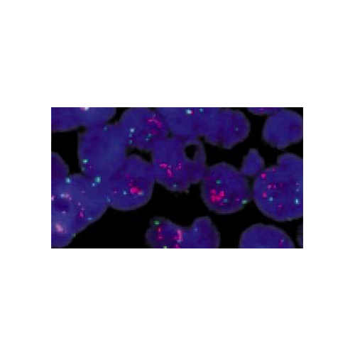
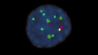
- Three alpha-satellite repeat sequence probes; CEP 3 SpectrumRed, CEP 7 SpectrumGreen, and CEP 17 SpectrumAqua that hybridize to the centromere regions of chromosomes 3, 7, and 17, respectively. In addition, a unique sequence probe, LSI p16 (9p21) SpectrumGold, is included that hybridizes to the p16 gene at 9p21.
- This probe set is premixed in hybridization buffer.
See microscope filter recommendations
Indications and Limitations Of Use
Intended Use
The UroVysion Bladder Cancer Kit (UroVysion Kit) is designed to detect aneuploidy for chromosomes 3, 7, 17, and loss of the 9p21 locus via fluorescence in situ hybridization (FISH) in urine specimens from persons with hematuria suspected of having bladder cancer. Results from the UroVysion Kit are intended for use, in conjunction with and not in lieu of current standard diagnostic procedures, as an aid for initial diagnosis of bladder carcinoma in patients with hematuria and subsequent monitoring for tumor recurrence in patients previously diagnosed with bladder cancer.
Limitations
- The UroVysion Kit has been optimized for identifying and quantitating chromosomes 3, 7, and 17, and locus 9p21 in human urine specimens.
- The performance of the UroVysion Kit was validated using the procedures provided in this package insert only. Modifications to these procedures may alter the performance of the assay.
- The clinical interpretation of any test results should be evaluated within the context of the patient's medical history and other diagnostic laboratory test results.
- UroVysion assay results may not be informative if the specimen quality and/or specimen slide preparation is inadequate, e.g., the presence of excessive granulocytes or massive bacteruria.
- Technologists performing the UroVysion signal enumeration must be capable of visually distinguishing between the red and green signals.
- Positive UroVysion results in the absence of other signs or symptoms of bladder cancer recurrence may be evidence of other urinary tract related cancers, e.g., ureter, urethra, renal, and/or prostate in males, and further patient follow-up is justified. In a study conducted on patients with hematuria (see "Symptomatic Patients: Performance vs. Standard of Care" for details on this clinical study) 3 patients, whose initial bladder cystoscopy was negative, were subsequently diagnosed with renal cancer within 6 months of this initial study visit. All 3 of these cases were positive by UroVysion.
- If UroVysion results are negative but standard clinical or diagnostic tests (e.g., cytology, cystoscopy) are positive, the standard procedures take precedence over the UroVysion test. Although the UroVysion Kit was designed to detect genetic changes associated with most bladder cancers, there will be some bladder cancers whose genetic changes cannot be detected by the UroVysion test.
- Ta stage solitary tumors smaller than 5mm could not be detected by UroVysion FISH.23 UroVysion FISH results are dependent on the amount of tumor cells that are deposited on the slide.
CAUTION: United States Federal law restricts this device to sale and distribution to or on the order of a physician or to a clinical laboratory; use is restricted to, by, or on the order of a physician.
.jpg)